Special stains in histopathology serve the primary purpose of identifying specific components within tissues, such as cellular structure ,fibrosis, calcification, proteins, carbohydrates, pigments,microrganisms and other residual elements. These stains help pathologists visualize and differentiate various constituents within tissues by highlighting them in distinct colors, enabling the identification of diseases .
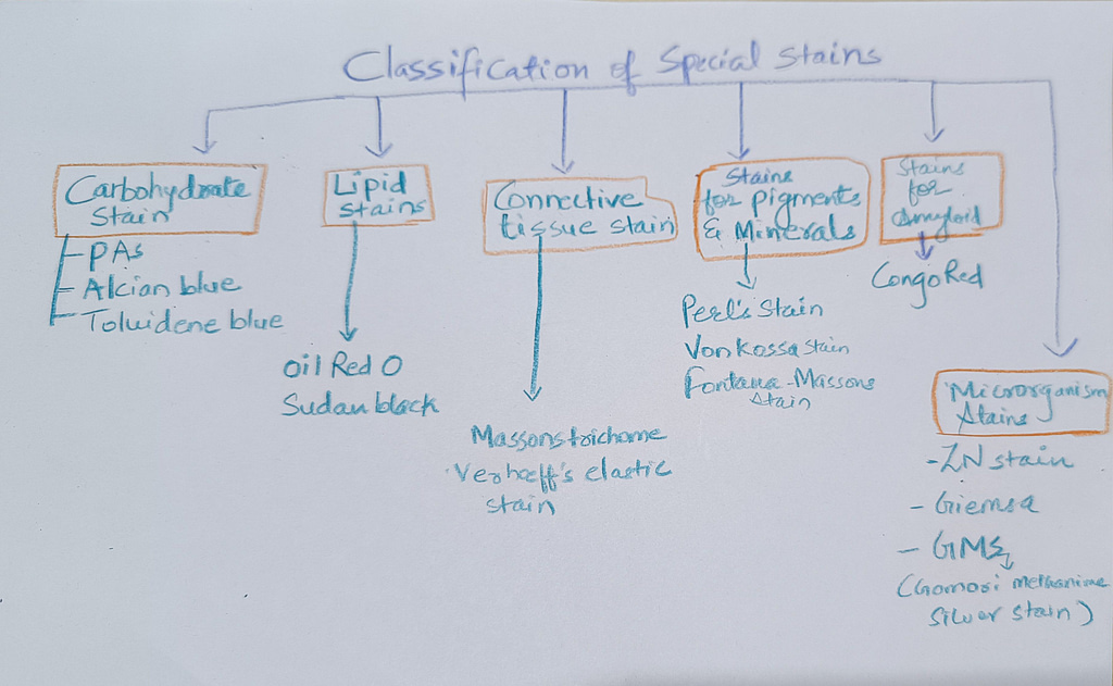
Classification The classification includes:
- carbohydrate stains
- Lipid stains
- connective tissue stains
- Stains for pigments and minerals
- Stains for Amyloid
- stains for microorganisms
Stains for carbohydrates
1PAS stain (Periodic acid-Schiff)
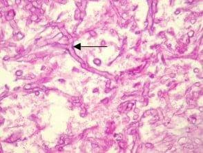
The dyes used in this technique involve 1% periodic acid and Schiff’s reagent. The substance which shows positive result to PAS has magenta color and nuclei are seen blue.The substances which show positive PAS are glycogen, neutral mucoprotein, glycoprotein, all fungi, cerebrosides, basement membrane.
Applications of PAS stain:
This stain is used in the diagnosis of several medical conditions
Tumors: mucins in adenocarcinoma of the large intestine, glycogen storage disorder, Ewing’s sarcoma, rhabdomyosarcoma, Whipple’s disease, etc
Demonstration of fungi in Fungal diseases like aspergillosis, mucormycosis,candidiasis
Renal biopsy:This stain is particularly valuable for its ability to highlight the basement membrane which is important in kidney transplant,diabetic nephropathy, amyloid etc
liver biopsy: PAS staining is particularly useful in detecting and evaluating glycogen storage disorders in liver tissues,and hepatitis, fibrosis, hepatocellular carcinoma.
2.Alcian blue
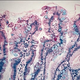
Blue staining is commonly employed to identify and characterize acidic mucin particularly those rich in sialic acid and carboxyl groups.
Application of Alcian blue: Diagnosis of barrets oesophagus, mucinous adenocarcinoma ,signet ring cell carcinoma
2.lipid stains
Oil Red O Stain for Fats and Lipids
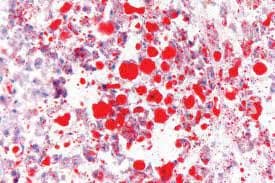
Oil Red O (‘ORO’) is used to demonstrate the presence of fat or lipids in fresh and frozen tissue sections, it stains neutral fat, fatty acids and triglycerides Routine tissue processing dissolves fat as it involves alcohol so its not possible to demonstrate fat in routine histology processing
Application of oil red O :
for diagnosing fat emboli in crush injury,diagnosing tumours with adipocytes origin
Sudan Black B: This stain is used to detect lipids in frozen sections, cytological preparations,also bone marrow and peripheral blood smear cells. It produces a black coloration in the presence of lipids.
3.Stains for connective tissue
Massons trichrome
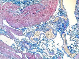
Masson’s trichrome stain combines the use of three dyes to selectively stain different tissue components. The technique involves staining nuclei and muscle fibers with Weigert’s iron hematoxylin, cytoplasm with Biebrich scarlet-acid fuchsin, and collagen fibers with aniline blue or light green. The staining process results in distinct colors for different tissue structures.
The nuclei appear dark blue or black, muscle fibers appear red, and collagen fibers are stained green or blue. This stain provides a sharp contrast between these components, enabling the identification and evaluation of tissues with collagenous structures, such as connective tissue, fibrotic tissue, and scar tissue.
Black-Nuclei
Red-Cytoplasm, muscle, erythrocytes
Collagen- blue
Application of Masson’s trichrome stain commonly used in the assessment of diseases involving fibrosis, such as liver fibrosis, myocardial fibrosis, and pulmonary fibrosis. It helps to visualize and quantify collagen deposition and fibrotic changes in tissues, aiding in the diagnosis and evaluation of various pathological conditions.
4. stain for pigment and minerals
perl stain
Perls stain, also known as Perl’s Prussian blue stain, is a histological staining technique used to detect iron deposits in tissue samples. It is commonly employed in the evaluation of iron-related disorders and pathological conditions.
Perls stain is based on the reaction between ferric iron and potassium ferrocyanide, resulting in the formation of Prussian blue, a blue-colored pigment. The stain selectively binds to iron, highlighting its presence in tissues.
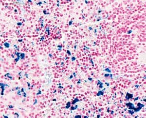
Blue colour is imparted to iron deposits(hemosiderin)
Application of perls stain;This staining technique is particularly useful in identifying iron accumulation in various tissues, including the liver, spleen, bone marrow. It aids in the diagnosis and evaluation of iron overload disorders such as hemochromatosis, as well as in detecting iron deposits in conditions such as hemosiderosis .When applied to tissue sections, Perls stain reveals blue-colored granules or aggregates, indicating the presence of iron. The intensity and distribution of the staining can provide information about the extent and pattern of iron accumulation within the tissue.
Masson Fontana Stain
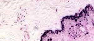
Used in identification of melanin, a pigment found in melanocytes and melanoma cells. It is also capable of detecting argentaffin granules, which are granules containing silver compounds like in carcinoid tumours.
Melanoma is a malignant tumor arising from melanocytes, the cells responsible for producing melanin. By staining melanin, the Masson Fontana method aids in the identification and evaluation of melanoma cells within tissue samples.This information is crucial for the diagnosis and evaluation of melanoma, helping to differentiate between benign nevi (moles) and malignant melanoma.
Van kossa stain for calcium
Van Kossa stain is a histological staining technique used to detect calcium deposits in tissue sections.Van Kossa stain utilizes the reaction between silver nitrate and calcium ions to form black-colored silver phosphate deposits. The stain selectively highlights calcium deposits, allowing for their visualization in tissue samples. useful in demonstration of pathological calcification like atherosclerosis
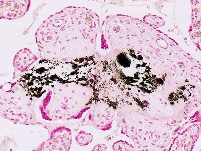
Calcium deposits stained with Van Kossa stain appear as black or dark brown areas within the tissue
5.Stain for Amyloid
what is amyloid?
This is an abnormally folded, fibrillar protein that deposits in extracellular
spaces in organs under certain pathological conditions. As it accumulates, it
progressively replaces the normal tissue of kidney,liver,brain,heart, and eventually results in loss of
function of vital organs, and ultimately, death.
Congo Red stain for amyloid
In H&E-stained tissue sections, amyloid, a proteinaceous material, may appear as an amorphous, glassy, eosinophilic substance. However, due to its resemblance to other materials, Congo Red (CR) staining is necessary to confirm its presence.
When CR-stained amyloid is observed under regular bright-field microscopy, it typically appears as a pale orange-red color. However, relying solely on the bright-field appearance is not sufficient for definitive amyloid identification, as small deposits may be challenging to detect. Therefore, CR-stained tissue sections must be examined under polarized light, enabling the visualization of a characteristic “apple green” birefringence that confirms the presence of amyloid.
The use of polarized light is essential because amyloid fibrils exhibit a unique property known as birefringence
Microorganism stains: These stains are used to visualize microorganisms in tissues. They are often used to diagnose infections, such as tuberculosis and leprosy. Some common microorganism stains include the Ziehl-Neelsen stain, the Gram stain, and the Giemsa stain.
