
Definition of Fixation: It is process in which cells or tissue are preserved ( fixed) in physical state and partly in chemical state so that they will with stand subsequent treatment with various reagents with a minimum loss, distortion or decomposition.
mechanism of fixation– stabilization and denaturation of proteins is the mechanism by which most of the fixatives provide mechanical strength to withstand tissue processing.
why fixation is required for tissue processing
- Prevention of tissue autolysis and bacterial attack
- To maintain the shape and volume of the tissues during subseguent procedures (e.g. clearing, embedding,etc.)
- To keep tissues as close to their natural state and to minimize change of natural color and appearanceTo prevent loss of tissue substances or rearrangement of tissue ingredients
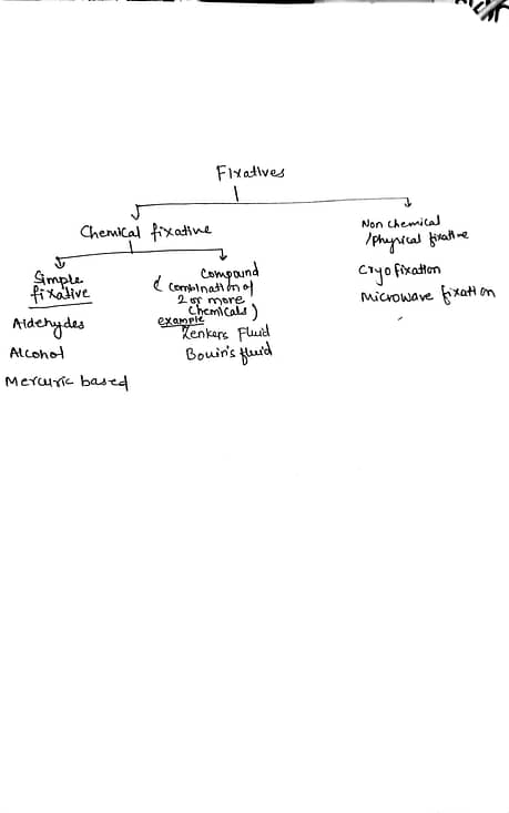
TYPES OF FIXATIVES
Fixatives can be classified based on their chemical composition and mode of action. fixatives are broadly grouped into Chemical and non chemical /physical fixatives,further chemical fixatives are typed on the basis of composition into simple and compound
- Aldehyde-Based Fixatives: Aldehyde-based fixatives are the most commonly used fixatives in histopathology.
- principle of fixation:They work by cross-linking proteins and preserving tissue structure. The primary aldehyde used in fixatives is formaldehyde. Examples of aldehyde-based fixatives include:
- Buffered 10% Formalin: most widely used in routine histopathology. It is a solution of formaldehyde gas dissolved in water and buffered to a slightly acidic pH, typically around 7.2 to 7.4. The buffering agents, such as phosphate salts, help maintain the pH stability of the solution.Formalin fixatives are versatile and applicable to various tissues and organs. They are particularly useful for routine histopathological examinations, as well as immunohistochemical and molecular studies.
- Bouin’s Fixative: Contains a mixture of formaldehyde, picric acid, and acetic acid, suitable for delicate tissues.Useful in demonstration of glycogen. Disadvantage it imparts yellow colour to tissue,reduces ferric content in tissue and makes tissue brittle if fixation done for more than 12 hours
- Glutaraldehyde :Glutaraldehyde fixation is extensively used in electron microscopy to prepare specimens for high-resolution imaging of cellular structures. It allows detailed visualization of organelles, membranes, and macromolecular complexes, providing valuable insights into cellular morphology and organization.
- Alcohol-Based Fixatives: Alcohol-based fixatives are used for rapid fixation and dehydration of tissues. These fixatives work by denaturation of proteins. They are commonly employed in frozen sections and cytological preparations. Examples include:
- Ethanol: Used for rapid fixation and preservation of cellular morphology in cytology,Hematology.
- Methanol: Particularly useful in cytological preparations and preservation of cellular components.
- They are commonly employed in frozen sections and cytological preparations. These fixatives are advantageous when immediate processing is required, but they may cause tissue shrinkage and lead to the loss of certain cellular components like lipid,myelin.
- Mercuric-Based Fixatives: Mercuric-based fixatives have declined in usage due to safety concerns regarding mercury exposure. However, they are known for their preservation of cellular morphology and antigenicity. Examples include:
- B-5 Fixative: Contains mercuric chloride and is used for immunohistochemical studies and electron microscopy
- Zenker’s Fixative: A mixture of formaldehyde, mercuric chloride, and potassium dichromate, used for preserving nuclear and cytoplasmic details.It is commonly used for the preservation of delicate structures, such as peripheral nerves and lymph nodes
Non-Chemical Fixatives (also called as physical fixatives)
Cryofixation: Rapid freezing of tissues using liquid nitrogen or carbon dioxide to preserve ultrastructural details.
Principles of Cryofixation: Cryofixation involves the rapid cooling of biological samples to extremely low temperatures, typically below -140°C (-220°F). The rapid freezing process minimizes the formation of ice crystals, which can cause damage to cellular structures.
Cryostat is freezing microtome that combines the functions of a microtome (instrument for cutting thin sections) and a freezing unit.Cryostats are used to cut thin frozen tissue sections for immediate examination during surgeries. This enables rapid intraoperative diagnosis and determination of margins during tumor excisions.

Two primary methods of cryofixation are commonly employed:
1.Liquid Nitrogen: In the liquid nitrogen method, the sample is directly immersed in or sprayed with liquid nitrogen. The extreme cold rapidly freezes the sample, preserving the cellular architecture.
2.High-Pressure Freezing: High-pressure freezing involves subjecting the sample to high pressure before freezing it. This method allows for the preservation of larger samples with minimal ice crystal formation.
Microwave-based Fixatives:
- Microwave fixation utilizes microwave energy( non-ionizing radiation) to accelerate the fixation process, reducing the overall turnaround time in histopathological studies .Microwave fixation allows rapid fixation and antigen retrieval, particularly useful for immunohistochemical studies.
- Principles of Microwave Fixation: Microwave fixation occurs through the interaction of microwave energy with tissues to achieve rapid fixation. Microwave radiation generates heat through dielectric heating, causing water molecules in the tissues to vibrate and generate thermal energy. This localized heating effect leads to the denaturation of proteins, cross-linking of macromolecules, and subsequent preservation of cellular structures.
Application of microwave fixation
- Immunohistochemistry (IHC): Microwave fixation is particularly advantageous for preserving antigens, making it suitable for immunohistochemical studies. It enhances antibody binding to target antigens, leading to improved staining results.
- Frozen Sections: Microwave fixation can be used in conjunction with frozen section techniques. It allows for rapid fixation of frozen tissue samples, ensuring preservation of cellular morphology and antigens prior to further processing and staining.
- Molecular Studies: Microwave fixation has been utilized in molecular pathology, including applications such as DNA, RNA, and protein extraction. It offers rapid and effective preservation of nucleic acids and proteins, facilitating downstream molecular analyses
Thank you for your information.
This detailed information helped me much in understanding the fixative and fixation process topic. Also the way you easily explained about microtome was best as I understood it perfectly.
Regards.