


MICROSCOPY
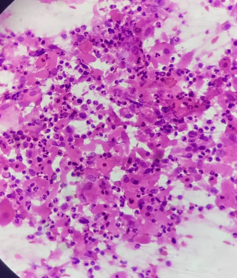
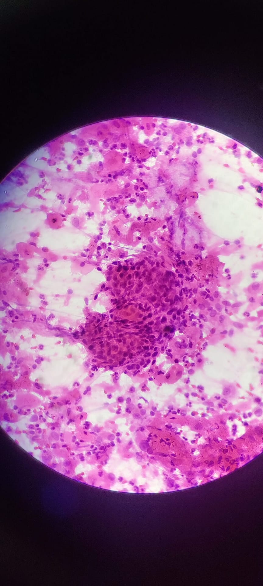
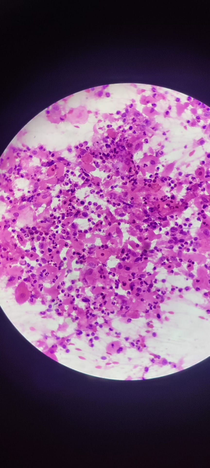
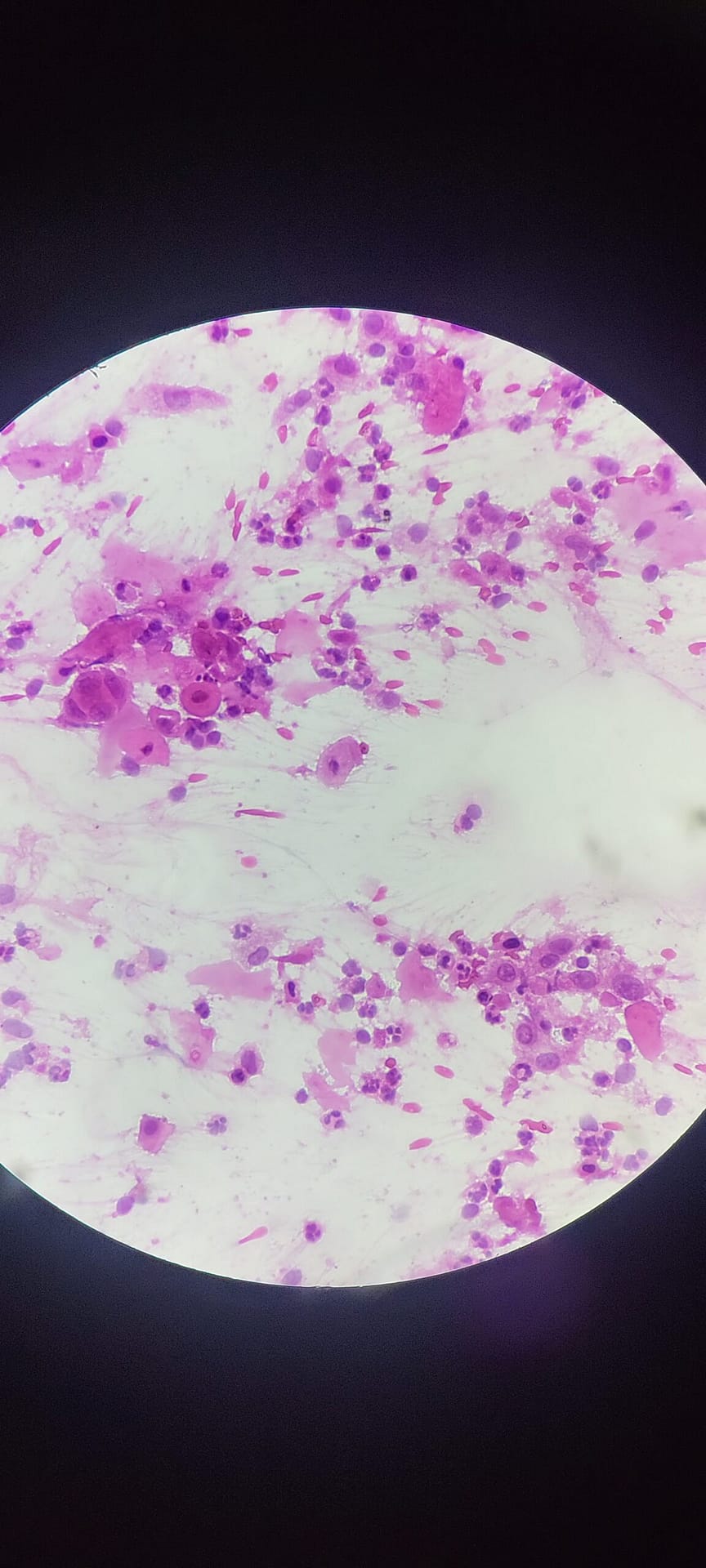
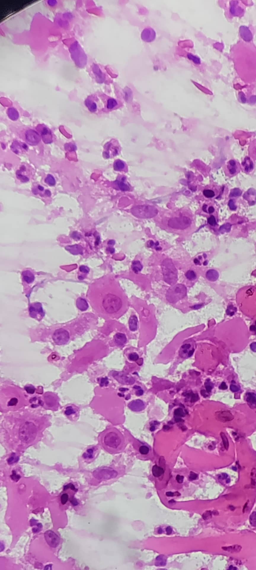
The smears of squamous cell carcinoma or metastatic Squamous cell carcinoma show clusters and sheets of malignant squamous cells accompanied by mature looking squamous epithelial cells. The cells exhibit marked pleomorphism, hyperchromasia, and irregular nuclear contours. Numerous keratinized and non-keratinized squamous cells are observed. Some cells display prominent nucleoli. The nuclei show irregular chromatin distribution, nuclear enlargement, and hyperchromasia. Cytoplasmic keratinization is evident in some cells.
Numerous atypical mitotic figures are noted, indicative of high proliferative activity. The background shows inflammation and necrotic debris.
In non keratinising variants ,orange cytoplasmic color is not seen. p40 stain helps in diagnosis of SCC.