In hematology, special stains are used to highlight specific cellular structures, components, or substances within blood and bone marrow samples. These stains provide additional information and aid in the identification of various cell types and abnormalities. Here are some commonly used special stains in hematology
Myeloperoxidase stain
The MPO stain, also known as Myeloperoxidase stain, is a special staining technique used in hematology to identify and highlight myeloid cells and their precursors. It specifically targets the myeloperoxidase enzyme present in these cells.
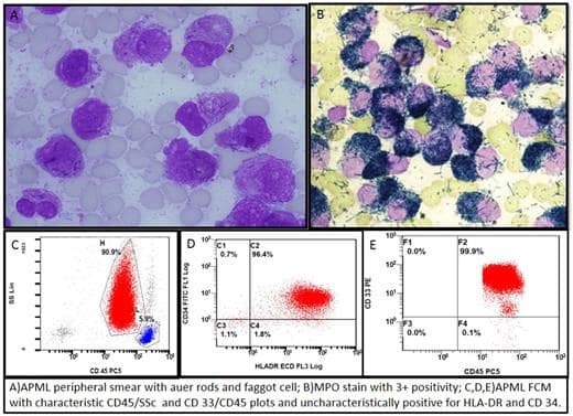
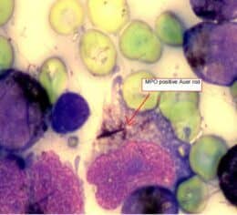
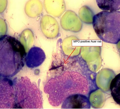
When applied, the MPO stain causes myeloid cells to exhibit a characteristic dark brown or blue-black coloration, making it easier for pathologists and hematologists to distinguish them from other cell types. This stain is especially useful in the diagnosis of acute myeloid leukemia (AML) and other related myeloid disorders.
Principle of MPO stain
The MPO stain utilizes hydrogen peroxide to activate myeloperoxidase in leukocyte granules. This enzyme oxidizes colorless substrates like benzidine, 3,3′-diaminobenzidine, or p-phenylenediamine dihydrochloride, resulting in the formation of an insoluble blue/brown derivative. The colored precipitate accumulates at the site of myeloperoxidase activity, enabling the identification of myeloid cells and their precursors in blood and bone marrow samples for diagnostic purposes.
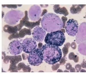
MPO stain causes myeloid cells to exhibit a characteristic dark brown or blue-black coloration
Uses of MPO stain: used for differentiating Acute Myeloid Leukemia from Acute Lymphoid Leukemia
preparation of MPO stain
| Ingredients | measurement |
| 30% Ethanol – | 100ml |
| Benzidine Dihydrochloride – | 0.3gm |
| 0.132 M Zinc Sulphate (3.8% W/V) | 1ml |
| Sodiun Acetate Trihydate- | 1gm |
| 3% Hydrogen Peroxide | 0.7ml |
| 0.1N Sodium Hydroxide (4.0% W/V)- | 1.5ml |
- The reagents are added in a beaker wrapped with aluminum foil in the given order only by mixing well after each addition.
- Benzidine salt may contain a small amount of inert non -soluble residue which gets dissolved upon addition of sodiumacetate.
- A precipitate will form when the zinc sulphate is added which gets dissolved after adding sodium acetate.
- The final pH should be 6.00 +/- 0.05.
- The solution should be filtered & kept at room temperature ina brown colored bottle, which is wrapped in aluminum foil.
- This solution is stable at room temperature and can be used
repeatedly for months
Counterstain
0.2% wright’s stain (2mg in 100ml of methanol)
Preparation of Sorensen’s buffer (pH 7.2, 0.15 M) for dilution;
a) KH2PO4 – 20.7 g/L- 20 ml
b) Na2HPO4-21.6 g/L- 80 ml
To obtain a solution of the required pH of 6.8, 20ml of (a) + 80ml of (b) is mixed.
PROCEDURE OF MPO STAINING
1.Fix air dried Peripheral blood smears or bone marrow slides in the fixative for 1 min
2.Wash in running tap water for 1 minute. Shake off excess water & blot with tissue paper gently. Dry
3.Place the slide in incubation mixture in Coplin jar for 30 seconds
4.Wash the slides in running tap water for 2 minutes (Yellow bluish green color should be completely washed out).
5.Counter stain with Wright’s stain and immediately dilute with Sorensen’s buffer. Keep it for 10 minutes.
6.Wash the slides in running tap water for 1 minute. Dry the slides and observe.
NOTE: MPO enzyme is sensitive to light and heat and hence the staining should be performed immediately or protected from light and heat.
Quality control
Positive controls : Myeloid series cells provide internal control.
Negative control: Internal control is provided by lymphocytes
Interpretation
- Myeloperoxidase is present in the primary granules of myeloid cells- promyelocytes,myelocytes, metamyelocytes, band form,neutrophils,eosinophils, weak in monocytes
- In myeloid series early myeloblasts are negative, with granular positivity appearing progressively as they mature.
- MPO is positive in AML-M1, M2, microgranular M3( Auer rods are strongly MPO positive).
- Cells of monocytic series display a less intense positive reaction that is characterized by fine granular deposits scattered throughout the cell.
- Lymphoblasts and lymphoid cells are MPO negative.